- DL manuals
- National
- Microscope
- 167 Series
- Instructions For
National 167 Series Instructions For
Summary of 167 Series
Page 1
National optical & scientific instruments inc. 6508 tri-county parkway schertz, texas 78154 phone (210) 590-9010 fax (210) 590-1104 instructions for 167, 168, 169, dc20-169, and 169-btw1 series compound biological microscopes copyright © 11/4/2016 national optical & scientific instrument inc..
Page 2
2 table of contents introduction / unpacking 167, 168 and 169 ............................................................................................... 3 assembly ......................................................................................................................................
Page 3
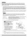
3 introduction thank you for your purchase of a national microscope. It is a well built, precision instrument carefully checked to assure that it reaches you in good condition. It is designed for ease of operation and years of carefree use. The information in this manual probably far exceeds what yo...
Page 4
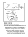
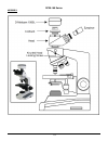
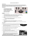
4 assembly a. Abbe condenser: pre-mounted in unit. B. Specimen holder: pre-mounted on stage. C. Objectives: unless otherwise note, they are pre-mounted on the microscope. D. Heads: on all series, the head comes pre-mounted to the body of the microscope. Position head so that it faces either forward ...
Page 5
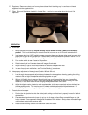
5 e. Eyepieces: remove the dust caps from eyepiece tubes. Avoid touching any lens surface and insert eyepieces into the eyepiece tubes. F. Filter: swing out filter holder has built in frosted filter – must be in place when using the 4x and 10x objectives. Operation g. Illumination. 1. Before operati...
Page 6
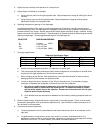
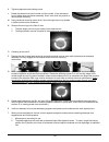
6 4. Adjust fine focus controls until specimen is in sharp focus. 5. Adjust diopter for difference in eyesight. A. Using right eye, peer into the right eyepiece tube. Adjust sharpness of image by utilizing fine focus controls. B. Using left eye, peer into the left eyepiece tube. Adjust sharpness of ...
Page 7
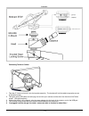
7 installing c-mount camera (to trinocular model only) trinocular model #169 is equipped with a port on top of head. Using the included c-mount adapter and the three-position sliding rod (c) allows use to easily direct microscope image into desired path. 1) rod pushed completely into head; 100% of m...
Page 8
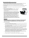
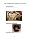
8 b. Electrical maintenance warning: for your safety, turn switch off and remove plug from power source outlet before maintaining your microscope. 1. Replacement of lamp. A. Carefully lay instrument on its side, taking care to avoid damage to the specimen slide holder located on top of mechanical st...
Page 9
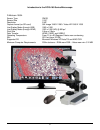
9 introduction to the dc20-169 series microscope d-moticam 1080l sensor type cmos sensor size 1/2.8" resolution 2 mp capture format (on sd-card) still image 1980 x 1080 / video hd 1980 x 1080 live display mode (through usb) 1980 x 1080 live display mode (through hdmi) 1920 x 1080 (hd) @ 60 fps* pixe...
Page 10
10 dc20-169 series assembly.
Page 11
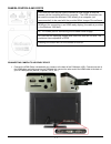
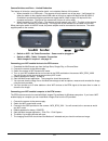
11 camera controls and ports connnecting camera to hd ready device 1. Connect the hdmi cable, included with your camera, to the back of the d-moticam 1080. Connect one end of the hdmi cable (end with screw) to the 1080 and then connect the other end of the hdmi cable to the back of your hd ready dev...
Page 12
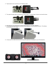
12 2. Plug the power cord into the back of the d-moticam 1080. 3. Then press the power button one time to turn on the camera. The green power indicator should now illuminate. 4. Now make sure your microscope is on and you are focused in on your slide / specimen. 5. On the right hand side of the head...
Page 13
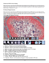
13 d-moticam 1080 on-screen display when the camera is connected to an hdmi display and controlled through a usb mouse, you can simply move the mouse cursor to the right to activate the on-screen function display for capturing images and also for adjusting various aspects of the image parameters. Yo...
Page 14
14 image adjustment controls ae/awb menu allows you to adjust: exposure, gain, gamma, brightness, light frequency, white balance, and color temp. Settings menu allows you to adjust: saturation, contrast, gamma, sharpness, digital noise, save individual user settings, time setting, language, format t...
Page 15
15 transfering images to your computer by default all your images are saved to an sd card, purchased separately. If you do not own one already, it is recommended you also purchase an external sd card reader. This is simplest and easiest way to transfer images to your computer. Images are automatical...
Page 16
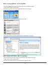
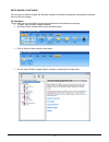
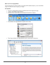
16 motic live imaging module – pc full help menu the full live imaging module manual is accessible within the live imaging main page. To begin, open the motic images software. At the top of main screen find the menu tab labeled file and click on capture: once the motic live imaging module has opened...
Page 17
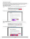
17 this will open the motic images help manual:.
Page 18
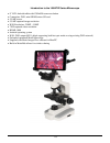
18 introduction to the 169-btw series microscope 8" lcd android tablet with 1280x800 screen resolution connection: wifi; mini hdmi; micro sd card 5.0 mp sensor 5.0 mp captured image resolution wifi resolution: 2.0mp - 5.0mp 720p captured video resolution hdmi: 1080 android operating system wifi: wif...
Page 19
19 assembly connecting tablet to camera 1. Take the 8” tablet and mount it into the bracket assembly. The bracket will hold the tablet at opposite corners of the tablet screen. 2. Taking the spiral usb cable cord and plug one end into your camera and the other into the side of the tablet screen. (se...
Page 20
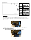
20 tablet operation power management power to the btw camera is supplied via the coiled usb cable provided with this product. Since the tablet batteries must supply power for both the tablet and camera simultaneously, approximately 2hrs of continued used can be expected. 8hrs of recharging is requir...
Page 21
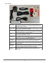
21 tablet accessories hdmi cable this cable will enable you to connect to an hdmi ready device, to view your tablet on a larger format. The displayed image will replicate the displayed image of the tablet. Power adapter this 5v dc power adapter is for charging your tablet’s battery. It is also used ...
Page 22
22 camera switches and ports – limited production this feature is limited to a small production batch, not a standard feature of this camera. When the switch is in usb mode – the camera will only transmit one of two ways. It will transmit to either the tablet via the supplied coiled usb cable or thr...
Page 23
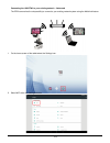
23 connecting the 169-btw1 to your existing network – advanced the btw camera has the unique ability to connect to your existing network system using the motichub feature. 1. On the home screen of the tablet select the settings icon. 2. Select wifi within the settings window..
Page 24
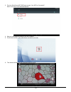
24 3. Find your work/home wifi ssid and connect. *turn wifi on if turned off* note: in this case i have selected national. 4. Now you have added the tablet to your wifi network 5. On the home screen of the tablet select the moticonnect app. 6. Then select the motichub icon and motichub will launch..
Page 25
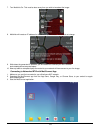
25 7. Turn motichub on. This must be done every time you wish to broadcast the image. 8. Motichub will create an ip address for the tablet on your network. This should not change. 9. Write down the generated ip address, you will need this to connect your wifi enabled device/tablet/phone/computer/lap...
Page 26
26 4. Click on the camera device button at the top right hand of the screen. 5. Click on the add button on the pop up window 6. You may give your new device any name, however the ip address must be same as the ip generated by motichub. 7. Now select your new camera.
Page 27
27 8. You can now view the image live on the moticonnect app as well as capture, measure and annotate the image using the tools provided. Connecting to networked btw with motic images 1. Install the motic images software into your computer or laptop. 2. Start the motic images application – click fil...
Page 28
28 4. The video device box should have the moticam x selected. 5. Click on open to open the moticam x ip address box. 6. In the open moticam x ip address box type in the ip address generated by the motichub feature of the btw tablet. 7. Once you click ok, the image produced by your btw tablet should...
Page 29
29 tablet software this android tablet comes equipped will all the necessary software needed to start using your equipment out of the box. Since it is an android tablet, you will be able to connect to your office/school wi-fi network to access the internet and download additional applications. The p...
Page 30
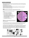
30 calibration – (*optional – not necessary to use your microscope*) to prepare for the calibration process, please make sure you have the calibration slide. The calibration slide has four individual dark round circles. Each dark round circle corresponds with each objective on your microscope. The 1...
Page 31
31 motic images 2.0 software you can use your transfer images with the motic images 2.0 software included with microscope to annotate, save and file your images. Full help menu the full software manual for motic images is accessible within the software’s main page. To begin, open the motic images so...
Page 32
32 motic live 2.0 live imaging module if you are connecting your camera via wifi to your laptop of wifi enabled computer, you can use the motic images 2.0 software to view and capture your images. Full help menu the full live imaging module manual is accessible within the live imaging main page. To ...
Page 33
33 motic images 3.0 software the motic images 3.0 software, like the motic images 2.0 software will allow you to view, capture, annotate and save your images. For further assistance in using the motic images 3.0 software please refer to the motic help files. These files will help explain the functio...
Page 34
34 model 926 phase contrast set introduction phase contrast microscopy provides a means to observe transparent specimens, which are very difficult to observe under bright field illumination. Another advantage of phase microscopy is that it allows the user to observe living specimens that are usually...
Page 35
35 k. Tighten eyepiece knurled locking screw. L. Rotate the phase turret annuli control until the number 10 can be seen at front of phase turret condenser assembly. Annuli must click into position to assure proper centering. M. Using condenser focusing control knob, focus the bright annuli ring loca...
Page 36
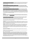
36 cleaning your microscope national microscopes are designed to function with minimal maintenance, but certain components should be cleaned frequently to ensure ease of viewing. The power switch should be turned off or the microscope should be unplugged when not in use. Do not disassemble your micr...
Page 37
37 for objectives that work with immersion oil it is essential to clean them after each observation session. To clean use a cleaning cloth for lenses slightly dampened with a low graduation of alcohol. Proceed by cleaning the frontal objective lens (normally 100x-oil). It is important for those obje...
Page 38
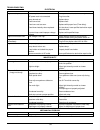
38 troubleshooting electrical problem reason for problem solution light fails to operate outlet inoperative. Ac power cord not connected. Lamp burned out. Fuse burned out. Fuse burns out too soon. Fuse blows instantly when replaced. Incorrect lamp used improper voltage or base. Have qualified servic...
Page 39
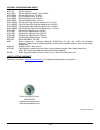
39 optional accessories and parts: #610-160 wf10x eyepiece #610-160r wf10x eyepiece w/reticle, 10mm/100div. #704-160sp din 4x objective lens, 0.10 n.A. #710-160sp din 10x objective lens, 0.25 n.A. #740-160sp din 40x objective lens, 0.65 n.A. #799-160sp din 100x objective lens, 1.25 n.A. #704-160asc ...

