- DL manuals
- OCULUS
- Medical Equipment
- Pentacam
- Interpretation Manual
OCULUS Pentacam Interpretation Manual
Interpretation Guide Pentacam
®
/ Pentacam
®
HR
InterpretatIon GuIde
3rd edition
Pentacam
®
Pentacam
®
HR
OCULUS
WWW.OCULUS.DE
64/09
15/EN/LA
P/SD/004/EN
OCULUS Optikgeräte GmbH
Postfach
•
35549 Wetzlar
•
GERMANY
Tel. +49-641-2005-0
•
Fax +49-641-2005-295
E-Mail: export@oculus.de
•
www.oculus.de
• OCULUS USA, info@oculususa.com
• OCULUS Asia, info@oculus.hk
• OCULUS Czechia, oculus@oculus.cz
• OCULUS Iberia, info@oculus.es
• OCULUS Poland, biuro@oculus.pl
• OCULUS Slovakia, office@oculus.sk
• OCULUS Turkey, info@oculus-turkey.com.tr
Summary of Pentacam
Page 1
Interpretation guide pentacam ® / pentacam ® hr interpretation guide 3rd edition pentacam ® pentacam ® hr oculus www.Oculus.De 64/09 15/en/la p/sd/004/en oculus optikgeräte gmbh postfach • 35549 wetzlar • germany tel. +49-641-2005-0 • fax +49-641-2005-295 e-mail: export@oculus.De • www.Oculus.De • o...
Page 2
Foreword we thank you for the trust you have put in us by purchasing this oculus instrument. In doing so you have chosen a modern, sophisticated product which was manufactured and tested according to strict quality standards. Our company has been doing business for over 120 years. Today oculus is a ...
Page 3
1 table of contents 1 introduction....................................................................................................................................................5 2 description of the unit and general remarks..........................................................................
Page 4
2 10 screening for refractive surgery by prof. Michael w. Belin................................................47-63 10.1 screening parameters, 4 maps refractive display......................................................................47 10.1.1 suggested installation settings ......................
Page 5
3 16 intacs® implantation...............................................................................................................117-122 16.1 case 1: by prof. Michael w. Belin....................................................................................................117 16.2 case 2: i...
Page 6
4 23 case reports from daily practice.............................................................................................161-172 23.1 case 1: cortical cataract by tobias h. Neuhann, md................................................................161 23.2 case 2: remove sutures after corne...
Page 7
5 1 introduction this guide is intended to assist pentacam®/pentacam® hr (referred to here as pentacam®) users in interpreting the results and screens of the pentacam®. We may not have covered everything which might be of interest, and we therefore ask anyone using the pentacam® for their help in im...
Page 8
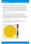
6 3 differences between the various topography maps of pentacam ® 3.1 calculation of corneal power corneal placido topographers measure geometrical corneal slope values. These values are converted into curvature values e.G. Values of axial (sagittal) curvature or instantaneous (tangential) curvature...
Page 9
7 c. The refractive index for historical reasons, most placido topographers and keratometers use the refractive index of 1.3375 for calculating corneal refractive power. However, this refractive index is actually incorrect even for the untreated eye (n ≈ 1.332). It assumes the ratio between the ante...
Page 10
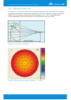
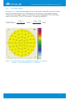
8 3.3 refractive power map this map (figure 3) uses only values from the anterior surface, but it also takes effect “a” (see above) into account. It calculates corneal power according to snell’s law of refraction assuming a refractive index of 1.3375 to convert curvature into refractive power (figur...
Page 11
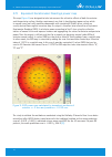
9 3.4 true net power this map (figure 4) shows the optical power of the cornea based on two different refractive indices, one for the anterior (corneal tissue: 1.376) and one for the posterior surface (aqueous humour: 1.336), as well as the sagittal curvature of each. These results are aggregated. T...
Page 12
10 the study to validate the method was conducted using the holladay 2 formula. Here it was deter- mined that after lasik the best correlation with the traditional method, with a mean prediction error of -0.06 d ± 0.56 d, is obtained using a mean zonal ekr for the 4.5 mm zone. For post-rk patients, ...
Page 13
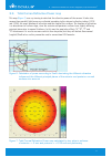
11 3.6 total cornea refractive power map this map (figure 7) uses ray tracing to calculate the refractive power of the cornea. It takes into account how parallel light beams are refracted according to the relevant refractive indices (1.376 and 1.336), the exact location of refraction and the slope o...
Page 14
12 4 recommended settings and color maps, displays and values 4 recommended settings and color maps, displays and values physicians who are starting to work with the pentacam® often turn to us with questions on settings such as step width on the color scale, or which maps and values to consider befo...
Page 15
13 4 recommended settings and color maps, displays and values 4.2 recommended color maps, displays and values 4.2.1 screening for corneal refractive surgery we recommend using the following maps and analysis displays: fast screening report to check whether the displayed parameters are within normal ...
Page 16
14 4.2.3 glaucoma screening we recommend using the following maps and analysis displays: fast screening report to check whether the displayed parameters are within normal limits general overview display to view the chamber angle in the scheimpflug images and corneal thickness. While clicking to the ...
Page 17
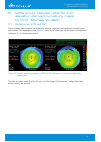
15 5 differences between placido and elevation-derived curvature maps by prof. Michael w. Belin 5.1 keratoconus in od and os? The case shown below explains the difference between suspicious and significant elevation maps and numbers. The topographic map (figure 8) shows the left and right eye but gi...
Page 18
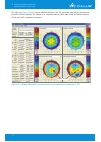
16 the right eye (figure 9) has a regular corneal thickness, but the elevation maps of the anterior and posterior surface indicates this cornea as a suspicious cornea. Both sides show an inferior position of the cone with suspicious elevations. Suspicious elevation figure 9: 4 maps selectable showin...
Page 19
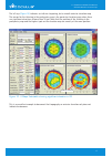
17 the left eye (figure 10) indicates an inferior steepening, but a smooth anterior elevation map. The reason for the thinning in the pachymetry map is the posterior elevation map, where there are significant elevations of more than 30 μm. Note that the position of the thinning in the pachymetry map...
Page 20
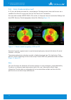
18 5.2 form fruste keratoconus? A 47-year-old female presented for a second opinion. She had previously been told she was not a candidate for refractive surgery and that she had “form fruste” keratoconus. Her exam had revealed a bscva 20/20+ od, and the slit lamp and external examination findings ha...
Page 21
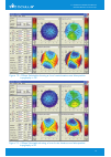
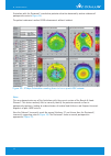
19 figure 12: 4 maps selectable showing a form fruste keratoconus false-positive topography in os figure 13: 4 maps selectable showing a form fruste keratoconus false-positive topography in od 5 differences between placido and elevation-derived curvature maps.
Page 22
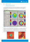
20 6 the fast screening report as a first step in examining a patient and evaluating one’s findings by ina conrad-hengerer, md the fast screening report is a very good way of gaining a quick overview when examining patients, especially when they are presenting for the first time. The pentacam® analy...
Page 23
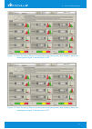
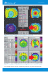
21 figure 14: fast screening report showing abnormal pachymetry and elevation data with unambiguous signs of keratoconus in od figure 15: fast screening report showing abnormal pachymetry and elevation data with unambiguous signs of keratoconus in os 6 the fast screening report.
Page 24
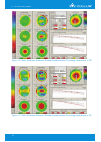
22 figure 16: belin/ambrósio enhanced ectasia display (version iii) showing keratokonus in od figure 17: belin/ambrósio enhanced ectasia display (version iii) showing keratokonus in os 6 the fast screening report.
Page 25
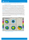
23 6.2 case 2: fuchs’ dystrophy with dmek cataract surgery – progress evaluation a 63-year-old female patient with bilateral cataract and fuchs’ dystrophy underwent combined cataract and dmek surgery. This section reports on her progress, documenting the condition of her right eye prior to surgery w...
Page 26
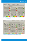
24 figure 19: fast screening report showing the presurgical condition in a case of fuchs’ dystrophy figure 20: fast screening report at one month after dmek surgery 6 the fast screening report.
Page 27
25 figure 21: corneal optical densitometry showing the presurgical condition in a case of fuchs’ dystrophy figure 22: corneal optical densitometry at one month after dmek surgery 6 the fast screening report.
Page 28
26 6.3 case 3: corneal injury sustained from an eye drop bottle after cataract surgery a 54-year-old patient underwent cataract surgery on his highly myopic right eye. The surgery was performed without any complications, resulting in a postoperative visual acuity of 20/20 with refraction values of s...
Page 29
27 figure 24: 4 maps refractive with suspicious curvature and elevation maps of the anterior surface figure 25: compare 2 exams showing changes in anterior surface elevation within a period of one week 6 the fast screening report.
Page 30
28 7 corneal power distribution display by ina conrad-hengerer, md 7.1 visual acuity impairment during nighttime driving with distance spectacles – nocturnal myopia? A driver had been wearing distance spectacles with the following refraction values for 2 years: od: sph -0.25 cyl -0.50 a 170° va 20/2...
Page 31
29 figure 26: 4 maps refractive showing with unremarkable elevation maps and curvature map in od figure 27: 4 maps refractive showing unremarkable elevation maps and curvature map in os 7 corneal power distribution display.
Page 32
30 figure 28: corneal power distribution showing normal power distribution in od figure 29: corneal power distribution showing a markedly increased power from 2.0 to 3.0 mm in os 7 corneal power distribution display.
Page 33
31 8 corneal ectasia 8 corneal ectasia 8.1 case 1: ectasia after radial keratotomy by prof. Renato ambrósio jr a 28-year-old male patient had rk (radial keratotomy) in 1995 for myopic astigmatism followed by rk enhancement three years later in os. Corneal topography was not performed prior to surger...
Page 34
32 8 corneal ectasia figure 31: 4 maps refractive of os showing post-lasik ectasia figure 32: pachymetry progression in od figure: 33 pachymetry progression in os the pachymetric progression is abrupt in both eyes, providing a significant indication of ectasia (figure 32, figure 33) . Probably mild ...
Page 35
33 8 corneal ectasia 8.2 case 2: ectasia after lasik? By prof. Michael w. Belin a 46-year-old female had undergone lasik 2 years prior. She was interested in further vision enhancement for her dominant right eye. Her best spectacle corrected visual acuity (bscva) was 20/20+ with sph -1.25 d. The ref...
Page 36
34 evaluation with the pentacam® revealed no posterior elevation abnormality and no evidence of postoperative ectasia (figure 35) . The patient underwent routine lasik enhancement without incident. Note: this case demonstrates one of the limitations with the current version of the bausch & lomb orbs...
Page 37
35 figure 36: orbscan ® 4 maps incorrectly suggesting ectasia in od figure 37, pentacam ® 4 maps selectable revealing there to be no post-lasik ectasia in this example orbscan® measured the pachymetry 37 microns (μm) thinner than pentacam®. 8 corneal ectasia.
Page 38
36 9 glaucoma 9 glaucoma 9.1 case 1: general screening by tobias h. Neuhann, md a 48-year-old white male patient presented for a second opinion on his glaucoma treatment. His father and grandfather had had glaucoma. After ten years of glaucoma medical treatment his ophthalmologist was now recommendi...
Page 39
37 9.2 case 2: yag laser iridectomy by eduardo viteri, md this is a 64-year-old female patient who was complaining of episodes of blurred vision and tearing. Her iop was 18 mmhg in both eyes. Her anterior chamber was shallow on slit lamp examination and her optic nerve had a cup/disc ratio of 0.6 in...
Page 40
38 after yag laser iridectomy had been performed several of her anterior segment measurements changed (figure 42) . This is quite evident in the differential display (figure 43) . Figure 42: general overview display 10 days after yag laser iridectomy in os showing improved aca and acd values figure ...
Page 41
39 the aca is 4º wider, and, although the acd only deepened 0.09 mm centrally, the main difference is evident in the periphery, where you can see changes ranging from 0.19 mm to 0.30 mm. This was enough to increase the acv from 64 to 92 mm³. Comments the pentacam® is quite useful for measuring the a...
Page 42
40 humphrey visual fields were full in both eyes (figure 47, figure 48) , and optical coherence tomography (oct) and retinal nerve fiber layer (rnfl) scans showed retinal thickness to be normal in both eyes (figure 49) . Figure 45: general overview display showing a low acv, shallow acd and narrow a...
Page 43
41 figure 47: 24-2 humphrey visual field: full visual field in od figure 48: 24-2 humphrey visual field: full visual field in os figure 49: spectral domain oct showingnormal rnfl thickness in both eyes 9 glaucoma.
Page 44
42 she underwent a prophylactic laser peripheral iridectomy in both eyes, following which acv increased from 64 to 94 μm, aca widened from 19.7 to 26.4 degrees and acd deepened from 1.83 to 2.08 mm. We previously demonstrated that a cutoff value of 113 mm3 for acv discriminates narrow angles with 90...
Page 45
43 figure 51, general overview display showing a low acv, shallow acd and narrow angle in os 9 glaucoma.
Page 46
44 humphrey visual fields revealed early defects in both eyes (figure 52, figure 53) , while the oct rnfl scan showed an abnormally thin rnfl corresponding to the visual field defects in both eyes (figure 54) . Figure 52: 24-2 humphrey visual field showing an early superior arcuate defect in od figu...
Page 47
45 9.4 evaluating the anterior segment in phacomorphic glaucoma by dilraj s. Grewal, md a 76-year-old caucasian female patient presented with acute pain and redness in her right eye. Her iop was elevated to 58, she had microcystic edema and her pupil was minimally reactive with her vision at “count ...
Page 48
46 figure 56: scheimpflug image showing increased acv, deeper acd and wider aca following removal of the lens and posterior chamber iol implantation 9 glaucoma.
Page 49
47 10 screening for refractive surgery 10 screening for refractive surgery by prof. Michael w. Belin 10.1 screening parameters, 4 maps refractive display 10.1.1 suggested installation settings the following are my guidelines for pre-operative refractive surgery screening for keratoconus: use the 4 m...
Page 50
48 10 screening for refractive surgery 10.1.2 proposed screening parameters it is essential to check the settings for the fitting zone of the bfs in the settings of the pentacam®, since this influences the borderline numbers (figure 57) . If you are using the manual (fixed) 9 mm zone for fitting the...
Page 51
49 10.1.3 strategy on how to go through the exams the way i usually go through the exams is: Î look at anterior elevation first Î look at posterior elevation Î look at the pachymetry and thickness distribution. Off-center distribution of corneal thickness is highly suspicious Î look at the symmetry ...
Page 52
50 this 4 maps selectable display (figure 59) shows a normal with-the-rule astigmatic cornea (astig. 2.6 d). Both the anterior and posterior elevations demonstrate a similar pattern, as does the anterior sagittal curvature. The curvature maps reveal a steep cornea (k1 = 47.6, k2 = 50.2), but the ele...
Page 53
51 this 4 maps selectable display (figure 60) shows a normal with-the-rule astigmatic cornea (astig. 4.1 d). Both the anterior and posterior elevations have a similar pattern, as does the anterior sagittal curvature. The anterior elevation map is symmetric, and the curvature shows a symmetric astigm...
Page 54
52 10.3 astigmatism on the posterior cornea this 4 maps selectable display (figure 61) shows only a small amount of anterior (astig. 1.1 d) but a larger amount of posterior astigmatism (a more defined astigmatic pattern). However, because the posterior cornea contributes a much smaller amount to the...
Page 55
53 10.4 spherical cornea this 4 maps selectable display (figure 62) shows a normal, relatively spherical cornea (astig. 0.7 d). The anterior elevation shows no defined pattern, which is mirrored by the anterior sagittal curvature. The corneal thickness is slightly high (583 μm in the thinnest readin...
Page 56
54 10.5 thin spherical cornea this 4 maps selectable display (figure 63) shows a relatively spherical anterior cornea (both anterior elevation and anterior sagittal maps) and a more pronounced astigmatic pattern on the posterior corneal surface. Because the optical properties of the posterior cornea...
Page 57
55 10.6 thin cornea this show 2 exams display (figure 64) shows from od and os the posterior elevation and the pachymetry maps. The posterior elevation shows a normal astigmatic pattern, as does the anterior elevation (not shown). The pachymetry maps show the thinnest regions od at 492 μm and os at ...
Page 58
56 10.7 borderline case of keratoconus this 4 maps selectable display (figure 65) shows a low-grade paracentral island (maximal elevation in island + 8 μm) in the anterior elevation map and a diffuse oval island on the posterior surface (maximal elevation in island + 16 μm). The anterior values are ...
Page 59
57 10.8 displaced apex this is a 4 maps selectable display of a normal astigmatic eye with a thick cornea (644 μm) (figure 66) . The anterior elevation map shows a "displaced apex" (displaced inferiorly). This causes the curvature map (anterior tangential curvature) to show an asymmetric pattern. Cu...
Page 60
58 10.9 pellucid marginal degeneration these are pictures of classic pellucid marginal degeneration (pmd). The pachymetry map (figure 69) shows the band of thinning located 1 - 2 mm from the inferior limbus. This is an area that cannot be imaged on a placido system, which is limited to imaging the c...
Page 61
59 10.10 asymmetric keratoconus this is a 4 maps selectable display (figure 70) of a normal astigmatic eye (od) with a thin cornea (thinnest reading 483 μm) and a noteworthy abnormality in the pachymetry distribution with a significant inferior-temporal displacement of the thinnest zone. At times th...
Page 62
60 the left eye shows a major posterior ectasia (+ 91 μm) on inferior island, and marked inferior displacement of the pachymetry map (thinnest reading 414 μm) (figure 71) . The anterior elevation shows a somewhat irregular astigmatic pattern but without any obvious positive island. The tangential cu...
Page 63
61 10.11 keratoconus with false negative findings on curvature map this 4 maps selecable display (figure 72) shows a classic keratoconus in os. The anterior elevation map shows a minor island that is still within the normal range. The posterior elevation, however, shows a very significant area of in...
Page 64
62 10.12 keratoconus greater in od than os look at the show 2 exams display of posterior elevation and pachymetry of od and os in this patient with keratoconus (figure 73) . The right eye shows a significant posterior island (ectatic area) associated with marked corneal thinning (430 μm) and signifi...
Page 65
63 10.13 classic keratoconus this 4 maps selectable display shows a case of classic keratoconus in od (figure 74) . Both anterior and posterior elevations show a prominent island of positive deviation (maximal at +33 μm anterior and +89 μm posterior) with an accompanying displacement of the pachymet...
Page 66
64 11 corneal thickness spatial profile by prof. Renato ambrósio jr the measurement of corneal thickness has become an important factor in a variety of clinical situations, including planning and evaluation of results of most types of corneal and anterior segment surgeries and evaluation of corneal ...
Page 67
65 percentage of increase in thickness (pit) pit can then be calculated for each position using the simple formula: Î clinical results in a published study involving 46 eyes of 23 patients (13 females) diagnosed with mild to moderate keratoconus and 364 normal eyes from 196 patients (97 females), st...
Page 68
66 it is worth noting that mandell and polse pioneered this field in a study using a modified haag- streit optical pachymeter with an electronic recording system to document the variation in thickness over the horizontal meridian measured at different angles [6] . However, this interesting approach ...
Page 69
67 11.1 screening for ectasia by prof. Renato ambrósio jr, marcela q. Salomão, md the new software combines the elevation criteria of michael belin, md for screening for ectasia. This opens up new horizons in analysing corneal thickness for diagnosis and classification of corneal ectasia. The ctsp a...
Page 70
68 figure 78: show 2 exams pachymetric showing a normal thin cornea figure 79: show 2 exams topometric showing an ectatic cornea 11 corneal thickness.
Page 71
69 figure 80: show 2 exams pachymetric showing an ectatic cornea currently, most diagnostic and classification criteria for keratoconus are based on anterior corneal curvature data derived from corneal topography. We wish to emphasize that the thickness profile described here should be used in conju...
Page 72
70 figure 81: show 2 exams topometric showing an asymmetric cornea figure 82: show 2 exams pachymetric showing an asymmetric cornea to test the hypothesis that the ctsp and pit increase sensitivity for the detection of very early forms of keratoconus we studied patients with keratoconus in one eye a...
Page 73
71 figure 83: show 2 exams topometric giving a false positive diagnosis of ectasia figure 84: show 2 exams pachymetric showing a normal cornea compared with the specificity of artificial intelligence based indices for detecting ectasia, topometric indices are fraught with high false positive rates, ...
Page 74
72 11.2 case 1: fuchs’ dystrophy by prof. Renato ambrósio jr, marcela q. Salomão, md contrary to ectasia, in which central thinning causes a more pronounced or abrupt increase in the thickness values from the center towards the periphery, corneal swelling makes the cornea homogeneously thick, decrea...
Page 75
73 figure 86: show 2 exams pachymetric showing a case of fuchs’ dystrophy 11 corneal thickness.
Page 76
74 figure 89: show 2 exams pachymetric showing thick corneas with abnormal corneal thickness progression in od and os figure 87: scheimpflug image showing clear cornea in od with no peak in the densitogram for the endothelium figure 88: scheimpflug image showing clear cornea in os with no peak in th...
Page 77
75 the iop, measured with the goldmann tonometer (2 p.M.), was 22 and 24 mmhg in od and os, respectively. Interestingly, the averages of two ocular response analyser measurements of goldmann- correlated iop, corneal compensated iop and hysteresis were 20.4, 17.8 and 13.2 mmhg in od and 25.1, 20.5 an...
Page 78
76 11.4 case 3: early fuchs’ dystrophy with glaucoma by prof. Renato ambrósio jr, marcela q. Salomão, md this 60-year-old patient was referred to us for a second opinion on his diagnosis of normal tension glaucoma, corneal disease and early cataract. The scheimpflug images show higher scatter (less ...
Page 79
77 figure 93: show 2 exams pachymetric showing an abnormal cornea in od and os the progression index was 0.5 in od and 0.8 in os, i.E. Lower than normal in both eyes. Goldmann iop (10 a.M.) was 18 mmhg in both eyes. The averages of two ocular response analyzer measurements of goldmann-correlated iop...
Page 80
78 completing the case report is the hrt examination displayed below. The optic nerve is damaged, and the rim configuration is abnormal both in the image and according to moorefield’s classification. This patient has glaucoma as well. Figure 94: specular microscopy in od and os figure 95: hrt single...
Page 81
79 11.5 screening parameters by prof. Renato ambrósio jr from my experience the following parameters can be used for screening corneal thickness profiles. It is very important to look out for blinking or significant fixation loss during the scan and repeat the exam whenever necessary. These paramete...
Page 82
80 12 belin/ambrósio enhanced ectasia display 12 belin/ambrósio enhanced ectasia display 12.1 why elevation is displayed by prof. Michael w. Belin before we can talk about how we display elevation tomographic data, we should take a step back and understand why i am a proponent of elevation based tom...
Page 83
81 the modern keratometers in use today are very similar to those used over a century ago and similar inherent limitations apply. The accuracy of the keratometer is conditional on the uniformity of the central corneal curvature over the area measured. The formula used by the keratometer assumes that...
Page 84
82 figure 98: keratoscope figure 99: lens form comparisons computerized videokeratoscopes provided a wealth of new information but still suffered from the same limitations of the century-old earlier techniques. Some of these limitations are related to the physical limits of reflective technology (pe...
Page 85
83 the pentacam® uses a technique of optical cross-sectioning to identify the anterior and posterior corneal surface, the anterior iris, and the anterior and posterior surface of the lens (figure 100) . By measuring these surfaces and their relative position elevation maps of the anterior and poster...
Page 86
84 the bfs provides the most clinically useful qualitative maps and is recommended for refractive screening, as conditions such as astigmatism and ectatic change are easily identified with it. While the bfs is the most clinically useful reference shape, it is important to realize that the normal cor...
Page 87
85 since the goal of refractive screening is to allow the physician to easily separate normal from abnormal, it works out that a bfs set at the 8.0 mm optical zone is optimal. A bfs computed from the central 8.0 mm will mask (i.E. Normalize) the normal asphericity and allow easy detection of patholo...
Page 88
86 most patients with keratoconus have in addition to a conical cornea a significant amount of astigmatism and one will typically see a positive island of elevation superimposed on an astigmatic pattern (figure 106) . While the bfs gives easily interpretable qualitative information, early or subtle ...
Page 89
87 the enhanced reference surface more closely resembles the more normal periphery (figure 108) and allows for easier identification of ectatic regions. In figure 109 , the standard bfs is shown on the left, while the enhanced reference surface on the right accentuates the ectatic region, yielding a...
Page 90
88 12.2 simplifying preoperative keratoconus screening by prof. Michael w. Belin, prof. Renato ambrósio jr, andreas steinmüller, msc the original version of the belin/ambrósio enhanced ectasia display changed the way we screened patients for sub-clinical ectatic disease. It was the first screening t...
Page 91
89 the combination of the pachymetric graphs and indices and elevation maps which utilize an enhanced reference sphere make possible an increased sensitivity and specificity in the screening of patients for ectatic disease. Each of these values (change in anterior elevation, change in posterior elev...
Page 92
90 figure 110: belin/ambrósio enhanced ectasia display (version ii) of a normal highly astigmatic eye both anterior and posterior elevations are normal and show a typical astigmatic pattern. The central cornea has a normal thickness at the apex of 532 μm but shows little progression towards the peri...
Page 93
91 figure 111: belin/ambrósio enhanced ectasia display (version ii) of a cornea with an isolated suspicious area on the posterior cornea the anterior elevation shows a very low degree of astigmatism, and the pachymetric progression, thinnest value and tp displacement are all within normal limits. Th...
Page 94
92 figure 112: interesting example of the value of the belin/ambrósio enhanced ectasia display (version ii) and the “d” values this cornea has a number of variables that fall in the suspicious area. Both the anterior and posterior elevations show small central yellow zones and the pachymetric progre...
Page 95
93 figure 113: belin/ambrósio enhanced ectasia display (version ii) of a case diagnosed as “mild” keratoconus based only on the anterior cornea. A fuller picture is obtained by tomography there are also changes on the posterior surface (both yellow), more dramatic changes in the pachymetric progress...
Page 96
94 12.3 interpretation of the belin/ambrósio enhanced ectasia display standard elevation maps: the left half of the belin/ambrósio enhanced ectasia display the elevation data is shown please refer to figure 113 . The first two elevation maps (placed side by side) display the baseline relative elevat...
Page 97
95 figure 114: belin/ambrósio enhanced ectasia display (version i) showing elevation data on the left and pachymetry data on the right) 12 belin/ambrósio enhanced ectasia display.
Page 98
96 12.4 pachymetry evaluation the pentacam® provides a detailed corneal thickness distribution map with 3 μm accuracy and repeatability. Display interpretation (pachymetry): the pachymetric portion of the display includes the pachymetry map (corneal thickness), the two graphs showing the current of ...
Page 99
97 figure 115: placido topopraphy in od showing no keratoconus figure 116: placido topography in os showing no keratoconus 12 belin/ambrósio enhanced ectasia display.
Page 100
98 had it only been judged on the basis of placido topography, cct and the clinical parameters, the case would have qualified as a good candidate for lasik. However, the pentacam® exam revealed some telling characteristics of the cornea which in our view constituted a high risk case for ectasia. Thi...
Page 101
99 figure 118: belin/ambrósio enhanced ectasia display (version i) showing subclinical keratoconus in od 12 belin/ambrósio enhanced ectasia display.
Page 102
100 12.6 early ectasia with asymmetric keratoconus by prof. Renato ambrósio jr, fernando faria-correia, md, allan luz, md a 20-year-old male patient with asymmetric keratoconus presented with bcva 20/20 in od and 20/15 in os. The data in os show a relatively normal topography pattern, while those in...
Page 103
101 figure 120: belin/ambrósio enhanced ectasia display (version iii) showing mild keratoconus in od figure 121: belin/ambrósio enhanced ectasia display (version iii) showing forme fruste keratoconus in os the belin/ambrósio enhanced ectasia display shows abnormal values for posterior elevation and ...
Page 104
102 12 belin/ambrósio enhanced ectasia display 13 locating the cone 13 locating the cone by prof. Michael w. Belin most clinicians have characterized keratoconus based on the appearance on curvature maps. This leads to inaccurate placement of the cone and a high incidence of supposed “pellucid margi...
Page 105
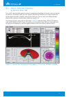
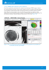
103 14 corneal optical densitometry display 14 the corneal optical densitometry display, by sorcha s. Ní dhubhghaill, mb, phd, jos j. Rozema, msc, phd gaining a sense of the structural situation in corneal pathologies can be difficult based on raw scheimpflug images alone. For this reason the method...
Page 106
104 14.1 keratic precipitates a 50-year-old patient presented with a history of granulomatous uveitis due to a toxoplasmosis infection. At initial presentation he had numerous large keratic precipitates deposited on the endothelial surface. In figure 123 the large precipitates are prominent on the i...
Page 107
105 figure 125: corneal optical densitometry display showing keratic precipitates after one week of therapy figure 126: corneal optical densitometry display showing keratic precipitates after two weeks of therapy 14 corneal optical densitometry display.
Page 108
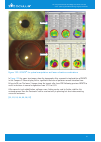
106 14.2 position and depth of intacs ® rings as described elsewhere in this interpretation guide, pentacam® examinations can be extremely useful both in planning for surgery and following up patients. The densitometry screen can add an extra dimension to the follow-up. A 35-year-old patient with a ...
Page 109
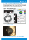
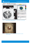
107 14.3 dsaek with specks at the interface one of the complications associated with a reduced visual outcome in dsaek surgery is haze at the interface between the donor cornea and the recipient cornea. The capacity of the densitometry screen to depict any desired layer makes it possible to examine ...
Page 110
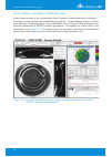
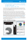
108 15 using pentacam ® technology to evaluate corneal scars, planning and documenting surgery outcomes by arun c. Gulani, md, ms i’d like to present the cornea as an elegant, optically powerful, and visually focused organ which is readily accessible for shape modification aimed at achieving unaided...
Page 111
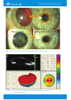
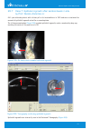
109 figure 130: post- and pre-op slit lamp images of an on-cornea scar figure 131: general overview showing findings on an in-cornea scar in the scheimpflug image 15 using pentacam ® technology to evaluate corneal scars, planning and documenting surgery outcomes.
Page 112
110 figure 132: pre- and post-op slit lamp images of an in-cornea scar the pentacam® corneal densitometry function is indispensable to me not only in selecting specific techniques but also as a means of educating my patients with a visual aid that they can understand. In cases of moderate anatomical...
Page 113
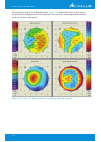
111 figure 133: intacs ® for optical manipulation and laser refractive combinations in figure 133 the upper two images show the topography after asymmetric implantation of intacs in the compare 2 exams display with a significant decrease of posterior corneal elevation from 84 μm to 28 μm. The lower ...
Page 114
112 15.1 case 1: corneal scar with rk incisions and cataract this patient was referred with a central on-corneal scar with multiple rk incisions and cataract. As a first step, he underwent scar peel with excimer laser myopic ablation to clear the cornea and make it measurable. After iol power calcul...
Page 115
113 figure 136: corneal optical densitometry display showing a clear cornea following laser scar peel; cataract present figure 137: slit lamp image showing a clear cornea following laser scar peel; cataract present 15 using pentacam ® technology to evaluate corneal scars, planning and documenting su...
Page 116
114 figure 138: corneal optical densitometry display showing status post cataract surgery with toric lens implant giving 20/20 vision figure 139: slit lamp image showing status post cataract surgery with toric lens implant giving 20/20 vision 15 using pentacam ® technology to evaluate corneal scars,...
Page 117
115 15.2 case 2: keratoconus with congenital cataract, high myopia and high astigmatism this patient was referred with keratoconus, congenital cataracts, high myopia, high astigmatism and presbyopia. As planned first stage, assymetric intacs® were implanted in preparation of lens exchange surgery us...
Page 118
116 figure 142: corneal optical densitometry display showing the emmetropic outcome after lens exchange surgery; intacs ® in place. Figure 143: slit lamp image after lens exchange surgery with an emmetropic outcome; intacs ® in place 15 using pentacam ® technology to evaluate corneal scars, planning...
Page 119
117 16 intacs ® implantation 16 intacs ® implantation 16.1 case 1: intacs ® implantation by prof. Michael w. Belin a 27-year-old female was referred by her optometrist because of poor vision od secondary to keratoconus. Her bscva was 20/200 od and with rgp over-refraction 20/30. The patient complain...
Page 120
118 16 intacs ® implantation then a complete pentacam® anterior segment analysis was performed, revealing the shortcomings of cone location and keratoconus classification based solely on anterior curvature. Both the anterior and posterior elevation map, as well as the pachymetry map locates the cone...
Page 121
119 16 intacs ® implantation 16.2 case 2: intacs ® after prk by alain-nicolas gilg, md a 45-year-old female had had photorefractive keratectomy (prk) in both eyes 7 years earlier. Before the laser surgery her visual acuity had been od: sph -7.50 cyl -0.50 a 170° os: sph -6.75 cyl -1.00 a 10° she was...
Page 122
120 figure 149: scheimpflug image after intacs ® implantation figure 148: pachymetric showing the pachymetry progression in an oblate postoperative cornea after the implantation of intacs® her visual acuity was os: sph +0.50 cyl -1.25 a 30° va 20/20 the scheimpflug image shows a successful fit of th...
Page 123
121 16.3 case 3: intacs ® & crosslinking by prof. Renato ambrósio jr, fernando faria-correia, md, allan luz, md a 24-year-old male patient presented with progressive keratoconus in os and anisometropia. Uncorrected visual acuity (ucva) was 20/20 in od and 20/200 in os. Wavefront assisted manifest re...
Page 124
122 16 intacs ® implantation figure 151: preoperative scheimpflug image of the vertical section figure 152: postoperative scheimpflug image of the vertical section.
Page 125
123 17 holladay report & holladay ekr65 detail report by jack t. Holladay, md 17.1 holladay report the holladay report was developed together with jack t. Holladay, md. The aim was: to improve the calculation of iol for patients who have undergone previous corneal surgery to design a display which c...
Page 126
124 then click “miscellaneous settings” to confirm all of the other settings. To keep these settings, select “holladay_setting” (figure 154) in the "load setting" pull-down menu, then click “save” and check the “lock settings” box. Figure 155 shows the map overlay for the corneal thickness map and f...
Page 127
125 figure 156: overlay for the 5 other maps the upper left box in figure 157 shows the general patient data. The center upper box in figure 157 shows the equivalent k-readings 65 (ekr65) [31] for the 4.5 mm zone along with mean ekr65, astigmatism, q-value (6 mm zone) and total spherical aberration ...
Page 128
126 the upper left map [axial/sagittal curvature (front)] in figure 157 , shows the axial curvature or power map uses a sph as the reference and the keratometric formula for power (337.5/axial radius in mm). A steel ball with a 7.5 mm radius of curvature would have a uniform power of 45.0 d (337.5/7...
Page 129
127 the central upper map (corneal thickness) in figure 157 , shows that the shape of a normal cornea is a negative meniscus lens (i.E., the back surface radius of curvature is steeper than the front) which is thinnest at its optical center and thickens by the square of the distance from the center....
Page 130
128 front elevation near min rp* elevation upper limit (μm) (μm) mean +1.0 -- +sd +2.1 +3.1 +2 sd +4.2 +5.2 +3 sd +6.2 +7.2 the lower right map [elevation (back)] (figure 157) shows the elevation of the back surface in μm relative to the bftef over an 8 mm zone. The eccentricity of the ellipsoid is ...
Page 131
129 17.2 holladay ekr65 detail report the upper box in figure 158 , shows the demographic information regarding the patient is in the box at the top of the page 2. The upper left box (figure 158) shows the table for the ekr65 (d) for various parameters from 1.0 to 7.0 mm [35,36,37] . All values are ...
Page 132
130 the lower central table in figure 158 , shows the ekr65 mean, is the weighted mean where 65% of the values are represented using the smallest range of points. In the above graph this value is 40.76 d (40.8 d) for the range indicated. Note that this value is less than the 40.93 d global mean of e...
Page 133
131 the ekr65 values at the top center (figure 159) are approximately 3.0 d flatter than average with 1.31 d of astigmatism. In the axial power map corneal power is steeper above than it is below by around 1.0 d, which is not unusual. The broken semi-meridian lines show mild irregular astigmatism wh...
Page 134
132 with a normal pupil of 5.48 mm the 4.5 mm grey column, with ekr65 flat k1 = 40.11 a 161° and steep k2 = 41.42 a 71° (figure 160) , would provide the correct keratometric values to enter into an iol calculator for the proper spheroequivalent power and toricity of the iol. An exact toric calculato...
Page 135
133 17.4 case 2: holladay report & holladay ekr65 detail report of a keratoconus exam the axial power map scale is centered at 43.0 d (green), which is normal. The axial and tangential curvature maps show a “hot spot” with the peak at 8 mm on the axial map and 6 mm on the tangential map (figure 161)...
Page 136
134 the distribution of ekr is a bimodal (2 peaks) (figure 162) distribution, and the ekr65 of 42.63 d is 0.57 d less than the 43.20 d peak. Standard keratometry usually measures nearer the peak and will over-estimate the power in keratoconus, leaving the patient with a hyperopic surprise. In severe...
Page 137
135 17.5 case 3: holladay report & holladay ekr65 detail report of a post lasik exam the mean value of the color scale (green) is 39.0 d, which is 4.5 d flatter than normal, and the semi- meridian lines are extremely segmented, demonstrating a large amount of irregular astigmatism. The ekr65 mean is...
Page 138
136 the ekr65 mean is 35.63 d at the 4.5 mm zone and 35.27 d at the 3.0 mm zone (figure 164) . The pupil diameter was 2.94 mm and confirmed with scotopic pupillometry done with another device. Due to the small pupil, the appropriate ks for iol calculation would be at the 3.0 mm zone and are ekr65 fl...
Page 139
137 18 corneal tomographic analysis is essential before cataract surgery - 4 steps in screening candidates for premium iols by prof. Naoyuki maeda new advancements in surgical techniques have improved the safety and efficacy of cataract procedures to a remarkable level. As a result, indications for ...
Page 140
138 18 corneal tomographic analysis is essential before cataract surgery - 4 steps in screening candidates for premium iols the pentacam® is a scheimpflug based corneal tomographer. We have been able to develop a program which makes it easy to perform the 4 steps of the screening procedure as descri...
Page 141
139 figure 166: cataract pre-op display moderate keratoconus figure 167: cataract pre-op display post lasik total hoa (1.575 μm) is high, and sa (-1.355 μm) is too low. Conventional spherical iol is recommended after obtaining the patient’s informed consent regarding the effects of corneal irregular...
Page 142
140 18.2 step 1: evaluation of corneal irregular astigmatism although there is no inherent problem in performing cataract surgery in patients with mild pterygium, subclinical keratoconus, or mild corneal scar, it is possible for irregular astigmatism associated with these corneal diseases to affect ...
Page 143
141 19 dependency of effective phacoemulsification time on pentacam ® nucleus staging (pns) 19 dependency of effective phacoemulsification time on pentacam ® nucleus staging (pns) by mehdi shajari md, wolfgang mayer md, prof. Thomas kohnen 19.1 introduction the pentacam® hr scheimpflug imaging syste...
Page 144
142 19 dependency of effective phacoemulsification time on pentacam ® nucleus staging (pns) 19.2 case 1: low pns and low ept in od of this caucasian female patient it can be seen by the relatively high scatter that the lens is not clear. Accordingly, the pentacam® shows a pns of 1. Please note also ...
Page 145
143 20 total corneal astigmatism for toric iol by giacomo savini, md the influence of posterior corneal astigmatism on total corneal astigmatism (tca) has been established by different studies based on scheimpflug imaging [45, 46, 47] . In the great majority of eyes, the steepest corneal meridian of...
Page 146
144 20.1 case 1: cylinder overcorrection from measurement of keratometric astigmatism in an eye with wtra a 51-year-old woman underwent cataract extraction in od. Her refraction was sph +4.00 cyl +0.75 a 100°. Axial length was 20.03 mm, and a +33.00 d toric acrysof (alcon, fort worth, tx) was calcul...
Page 147
145 the choice of the iol was between the sn6at3 and the sn6at4 models (the t2 model was not available). According to the manufacturer, the sn6at3 and the sn6at4 should correct, respectively, 1.03 and 1.55 d at the corneal plane. After including the effect of surgically induced astigmatism (0.2 d 90...
Page 148
146 20.2 case 2: cylinder undercorrection from measurement of keratometric astigmatism in an eye with atra an 85-year-old man underwent cataract surgery in os. Axial length was 23.31 mm, and a 21.50 d toric acrysof was calculated for emmetropia. Traditional ka measurement (figure 172) showed a value...
Page 149
147 20 total corneal astigmatism for toric iol after considering the sia = 0.2 d 90° the choice was between a sn6at4 to correct the ka (predicted residual astigmatism = 0.17 d 138°) and a sn6at6 to correct the tca (predicted residual astigmatism = 0.13 d 122°). The former should correct 1.55 d at th...
Page 150
148 20 total corneal astigmatism for toric iol we aimed to correct the tca and considering a surgically induced astigmatism of 0.2 d 90°, we targeted a correction of 1.37 d 76°. A sn6at4 iol was implanted at 75°. One month after surgery the refraction was sph -0.25 cyl -0.25 a 120°. Using the ka dat...
Page 151
149 21 overview about iol power calculation formulas for different eye types patient group iol-calculator/formula where to find value paper normal eyes standard historical methods (srk i; srk ii; hollady i; holladay ii; srkt; hoffer q; haiges and many others) cataract pre-op display sim k's and othe...
Page 152
150 22 phakic iol implantation 22 phakic iol implantation 22.1 manual pre-op simulation and post-op control by eduardo viteri, md 22.1.1 preoperative evaluation a high myopic, 20-year-old patient entered the office; refraction data: od: sph -12.00 (cyl -1.50 a 180°) os: sph -12.50 (cyl -1.50 a 10°) ...
Page 153
151 22 phakic iol implantation 22.1.2 postoperative evaluation the scheimpflug image shown below displays the same case after successful artisan piol implantation. It is evident there is space from the anterior piol surface to the endothelium centrally and at the periphery (figure 177) . The posteri...
Page 154
152 22 phakic iol implantation her pupil sizes were: scotopic: od 6.24 mm, os 6.27 mm mesopic low: od 4.74 mm, os 4.78 mm mesopic high: od 3.62 mm, os 3.56 mm. Because of her high ametropia, lasik and prk were not an option. Therefore we checked the possibility of a piol implantation. A 3d simulatio...
Page 155
153 22 phakic iol implantation because of these distances between the piol and the endothelium, it would be currently safe. The mimimum clearance between the piol and the endothelium was 1.22 mm calculated in 3d. The iris convexity is shown on the lower right hand side of figure 178 . However, what ...
Page 156
154 22 phakic iol implantation the image below shows the simulated piol position 20 years after implantation. The horizontal scheimpflug image shows a minimum clearance between optic edge and endothelim of 920 µm , but the distinct minimum clearance, derived from the internal 3d-model between the pi...
Page 157
155 22 phakic iol implantation 22.2.2 toric artisan/verisyse, 5/8.5 mm a 22-year-old female: od: sph -18.50 cyl 4.00 a 127° os: sph -10.50 cyl 1.50 a 45° asked for refractive surgery. On the left cornea a small corneal cicatrix was detected centrally during slit lamp examination. Her pupil sizes wer...
Page 158
156 22 phakic iol implantation since the patient was only 22 years old, we wanted to check the piol position as to be on the safe side. We checked the predicted piol position at 40 years after surgery (figure 182) . Here the simulated minimum clearance is 1 mm. Conclusion: the amount of endothelium ...
Page 159
157 22 phakic iol implantation 22.3 patient selection criteria by prof. Burkhard dick, sabine buchner, optometrist careful pre-op evaluation and patient selection are essential prerequisites for a successful implantation. There are several criteria that have to be considered. In our clinic, we look ...
Page 160
158 22 phakic iol implantation figure 184: 3d piol simulation and aging prediction showing a large iris convexity the following warning appears: “automatic piol alignment may be negatively influenced by large iris convexity”..
Page 161
159 22 phakic iol implantation 22.4 case example of ectasia after lasik, crosslinking and piol implantation by prof. Renato ambrósio jr, fernando faria-correia, md, allan luz, md a 25-year-old male patient presented with severe and progressive visual loss (ucva of counting fingers at 1 m in both eye...
Page 162
160 22 phakic iol implantation clinical case of post-lasik ectasia treated by transepithelial crosslinking followed by phakic intraocular lens implantation in both eyes. Figure 186: scheimpflug image after transepithelial crosslinking in od (a) and os (b), slit-lamp photograph showing piol implant i...
Page 163
161 23 case reports from daily practice 23 case reports from daily practice 23.1 case 1: cortical cataract by tobias h. Neuhann, md a 23-year-old, -12.5 d myopic white female underwent a fundus examination at a local eye clinic. Her bscva was 20/30, while her best corrected visual acuity with contac...
Page 164
162 23 case reports from daily practice 23.2 case 2: remove sutures after corneal transplant surgery? By tobias h. Neuhann, md a 22-year-old white male had received a corneal transplant due to keratoconus 12 months earlier. The first suture had already been removed. Examination with the pentacam® re...
Page 165
163 23 case reports from daily practice 23.3 case 3: keratoconus and cataract by tobias h. Neuhann, md a 54-year-old male asked for glasses. Bscva was 20/80 in both eyes. The pentacam® delivered the solution in two seconds. The right eye had a cataract and undetected keratoconus (figure 190, figure ...
Page 166
164 23 case reports from daily practice figure 193: 4 maps refractive of os figure 194: pachymetry progression in os figure 195: scheimpflug image of os.
Page 167
165 23 case reports from daily practice figure 196: topography of the central part of the cornea our recommendation was: cataract surgery on the right eye; deep lamellar keratoplasty for the left eye; and toric iol implantation in the left eye after suture removal. This still left one question open ...
Page 168
166 23 case reports from daily practice3 23.4 case 4: corneal infiltrate by prof. Renato ambrósio jr a 33-year-old female presented for evaluation of a corneal infiltrate. The patient mentioned having used tap water for cleaning her soft contact lens two days ago. Her symptoms had started after wear...
Page 169
167 23 case reports from daily practice she had been prescribed topical drops containing a combination of neomycin, polymixin b and dexametasone, which she had used 4 times since the last night, obtaining mild improvement. The patient was advised to discontinue contact lens use in both eyes and aske...
Page 170
168 23 case reports from daily practice 23.5 case 5: incisional edema by prof. Renato ambrósio jr a 76-year-old female patient presented with incisional edema 12 months after phacoemulsification. Endothelial morphology revealed large cells with pleomorphism and polymegathism. Central cell count was ...
Page 171
169 23 case reports from daily practice a slit lamp exam and a pentacam® exam were performed, and the findings were correlated with each other (figure 201, figure 202) . The central cornea was clear with no edema. The peripheral cornea at the incision location was edematous, with formation of small ...
Page 172
170 23 case reports from daily practice figure 204: general overview display revealing corneal thinning a pentacam® exam is useful for documenting corneal thickness. The thinnest spot is displayed in the pachymetry map and can also be seen in the scheimpflug images, facilitating follow-up examinatio...
Page 173
171 23 case reports from daily practice 23.7 case 7: epithelial ingrowth after keratomileusis in situ by prof. Renato ambrósio jr a 41-year-old male patient with a history of in situ keratomileusis in 1991 and one re-treatment for removal of epithelial ingrowth asked for a second opinion. The slit l...
Page 174
172 23 case reports from daily practice figure 207: part of 4 maps refractive confirming epithelial ingrowth the pachymetry map in the 4 maps refractive (figure 207) showed this effect as well, even in the presence of an opaque cornea. The pentacam® was useful for evaluating corneal elevation, curva...
Page 175
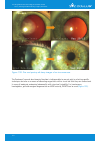
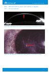
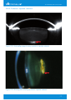
173 24 scheimpflug and slit lamp images 24 scheimpflug and slit lamp images 24.1 corneal dystrophy figure 209: slit lamp photo documenting corneal dystrophy on the posterior surface figure 208: scheimpflug image revealing, corneal dystrophy on the posterior surface.
Page 176
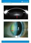
174 24 scheimpflug and slit lamp images 24.2 congenital anterior pyramid cataract figure 211: slit lamp photo showing an anterior pyramid cataract figure 210: scheimpflug image showing an anterior pyramid cataract.
Page 177
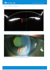
175 24 scheimpflug and slit lamp images 24.3 posterior capsular cataract figure 213: slit lamp photo showing a posterior capsular cataract figure 212: scheimpflug image showing a posterior capsular cataract.
Page 178
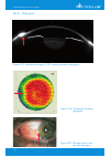
176 24 scheimpflug and slit lamp images 24.4 nuclear cataract figure 215: slit lamp photo showing a nuclear cataract figure 214: scheimpflug image showing a nuclear cataract.
Page 179
177 24 scheimpflug and slit lamp images 24.5 posterior synechia figure 217: slit lamp photo showing posterior synechia figure 216: scheimpflug image showing posterior synechia.
Page 180
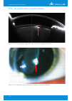
178 24.6 pterygium figure 219: topography revealing pterygium figure 220: slit lamp photo of an eye with pterygium figure 218: scheimpflug image in 190° showing a case of pterygium 24 scheimpflug and slit lamp images.
Page 181
179 25 orthokeratology, general screening by alain-nicolas gilg, md a 34-year-old male presented with the request to have his soft contact lenses changed because of progressive intolerance during the day. Subjective refraction resulted in a visual acuity of od: sph -2.50 os: sph -1.00 the pentacam® ...
Page 182
180 25 orthokeratology, general screening the patient was examined 4 times within 2 months to follow up the condition of the cornea, and the efficacy of the treatment was confirmed by a comparison of all four exams (figure 222) . Figure 222: compare 4 exams, one prior and three after orthokeratology.
Page 183
181 25 orthokeratology, general screening on day 28 the patient complained of fluctuations in his visual acuity during the day. Thereupon he was examined in the morning after wearing the lens over night and in the late afternoon. Examination with the pentacam® using the compare 2 exams display confi...
Page 184
182 26 important studies and case reports 26 important studies and case reports 26.1 refractive studies: 2014 a prospective study of pterygium excision and conjunctival autograft with human fibrin tissue adhesive: effects on vision, refraction, and corneal topography - (stuti misra, msc, boptom, jen...
Page 185
183 26 important studies and case reports 2012 quality of vision in eyes after selective lamellar keratoplasty – (shizuka koh, md, naoyuki maeda, md, tomoya nakagawa, md, and kohji nishida, md) - cornea _ volume 31, number 11, suppl. 1, november 2012 scheimpflug camera measurement of anterior and po...
Page 186
184 26 important studies and case reports corneal thickness changes after corneal collagen crosslinking for keratoconus and corneal ectasia: one-year results – (steven a. Greenstein, ba, vinnie p. Shah, md, kristen l. Fry, od, ms, peter s. Hersh, md) - j cataract refract surg. 2011; 37:691–700 q 201...
Page 187
185 26 important studies and case reports 2010 corneal volume, pachymetry, and correlation of anterior and posterior corneal shape in subclinical and different stages of clinical keratoconus – (david p. Pin˜ ero, msc, jorge l. Alio´ , md, phd, alicia aleso´ n, od, munir escaf vergara, md, mauricio m...
Page 188
186 26 important studies and case reports intra- and postoperative variation in ocular response analyzer parameters in keratoconic eyes after corneal cross-linking – (paolo vinciguerra, md; elena albè, md; ashraf m. Mahmoud, phd; silvia trazza, bs;farhad hafezi, md; cynthia j. Roberts, phd) - j refr...
Page 189
187 26 important studies and case reports anterior segment measurements using pentacam® and orbscan ii 1 to 5 years after refractive surgery – (sun woong kim, md; hae jung sun, md; jee ho chang, md; eung kweon kim, md, phd) - j refract surg. 2009;25:1091-1097 location of steepest corneal area of con...
Page 190
188 26 important studies and case reports repeatability and concordance of the pentacam® system. Comparative study of corneal parameters measured with pentacam® and atlas – (b. Doménech, d. Mas, e. Ronda, j. Pérez, j. Espinosa, c. Illueca) - optica pura y aplicada 2009; 42(1):51-60 2008 diurnal vari...
Page 191
189 26 important studies and case reports corneal curvature and central corneal thickness in eyes with pseudoexfoliation syndrome – (ibrahim f. Hepsen, md; ramazan yagci, md; urgcan keskin, md) - can j opthalmol 2007; 42:667-680. Doi:10.3129/can j ophthalmol.I07-145 graft central thickness measureme...
Page 192
190 intrasession and intersession repeatability of the pentacam® system on posterior corneal assess- ment in the normal human eye – (davie chen, andrew k.C. Lam, phd, faao) - j cataract refract surg. 2007; 33:448–454. Doi:10.1016/j.Jcrs.2006.11.008 keratoconus: it is hard to define, but – (michael w...
Page 193
191 26.2 case reports: corneal ectasia after lasik despite low preoperative risk: tomographic and biomechanical findings in the unoperated, stable, fellow eye – (renato ambrósio, jr, md, phd; daniel g. Dawson, md; marcella salomão, md; frederico p. Guerra, md; ana laura c. Caiado, md; michael w. Bel...
Page 194
192 scheimpflug analysis of corneal power changes after myopic excimer laser surgery – (giacomo savini, md, kenneth j. Hoffer, md, michele carbonelli, md, piero barboni, md) - j cataract re- fract surg. 2013; 39:605–610 q 2013 ascrs and escrs comparison of methods to measure corneal power for intrao...
Page 195
193 anterior chamber depth in normal subjects by rotating scheimpflug imaging – (matthew t. Feng, md a, michael w. Belin, md, franzco , renato ambrosio jr., md, phd, satinder p.S. Grewal, md, wang yan, md, phd, mohamed s. Shaheen, md, phd, charles mcghee, md, phd, naoyuki maeda, md, tobias h. Neuhan...
Page 196
194 clinical application of a scheimpflug system for lens density measurements in phacoemulsification – (jung-sub kim, md, so-hyang chung, md, phd, choun-ki joo, md) - j cataract refract surg. 2009; 35:1204-1209 anterior chamber depth measurement in pseudophakic eyes: a comparison of pentacam® and u...
Page 197
195 correlation of lens density measured using pentacam® scheimpflug system with locs iii grading score and visual acuity in age-related nuclear cataract – (xueting pei, yongzhen bao, yi chen and xiaoxin li) - br. J. Ophthalmol. Published online 27 jun 2008. Doi:10.1136/bjo.2007.136978 alterations i...
Page 198
196 in vivo measurement of opacified h60m intraocular lenses using scheimpflug photography – (a h ross, m v mundasad, s m neilson, e j mayer, j m sparrow, a d dick, d m tole) - br. J. Ophthalmol. 2006; 90;1328-1329 comparison of three methods of measuring corneal thickness and anterior chamber depth...
Page 199
197 2011 comparing corneal variables in healthy subjects and patients with primary open-angle glaucoma – (federico saenz-frances, julian garcia-feijo, luis janez, lara borrego-sanz, jose m. Martinez de la casa, ana fernandez-vidal, carmen mendez-hernandez, enrique santos-bueso, juan reche- frutosand...
Page 200
198 26.6 case reports: corneal ectasia after lasik despite low preoperative risk: tomographic and biomechanical findings in the unoperated, stable, fellow eye – (renato ambrósio, jr, md, phd; daniel g. Dawson, md; marcella salomão, md; frederico p. Guerra, md; ana laura c. Caiado, md; michael w. Bel...
Page 201
199 27 references 27 references 1. Tonn et al, anterior surface–based keratometry compared with scheimpflug tomography–based total corneal astigmatism; invest ophthalmol vis sci. 2015;56:291–298. Doi:10.1167/ iovs.14- 15659 2. Jack t. Holladay, md, msee, facs; warren e. Hill, md, facs; andreas stein...
Page 202
200 19. Ambrosio r jr, dawson dg, salomao m, et al. Corneal ectasia after lasik despite low preoperative risk: tomographic and biomechanical findings in the unoperated, stable, fellow eye. J refract surg 2010; 26(11):906-11 20. Ambrosio r jr, randleman jb. Screening for ectasia risk: what are we scr...
Page 203
201 38. Li wang, md, phd, douglas d. Koch, md. Custom optimization of intraocular lens asphericity. J cataract refract surg 2007; 33:1713–1720 39. Holladay jt. Exact toric iol calculations using currently available lens constants. Arch ophthalmol. Holladay jt 2012;130(7): 946-7 40. Martinez ce, klyc...
Page 204
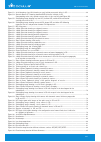
202 28 list of illustrations 28 list of illustrations figure 1: sagittal power map of a sphere, r = 8 mm.............................................................................................................................. 7 figure 2: snell´s law of refraction...................................
Page 205
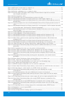
203 figure 53: 24-2 humphrey visual field showing an early inferior paracentral defect in os........................................................ 44 figure 54: spectral domain oct showing abnormal rnfl thickness inferiorly in od, corresponding to the early superior arcuate defect in that eye (als...
Page 206
204 figure 104: bfs-based elevation map of an astigmatic eye................................................................................................................... 85 figure 105: elevation map of a keratoconic cornea...........................................................................
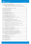
Page 207
205 figure 147: zernike analysis topography pre intacs®............................................................................................................................119 figure 148: pachymetric showing the pachymetry progression in an oblate postoperative cornea............................
Page 208
206 figure 196: topography of the central part of the cornea......................................................................................................................165 figure 197: scheimpflug image of a corneal infiltrate....................................................................
Page 209
207 29 tables directory table 1: central keratometry (front) (*n = 1243 normal eyes, internal, unpublished data by j. T. Holladay)................126 table 2: maximum tangential k (front) (*n = 1243 normal eyes, internal, unpublished data by j. T. Holladay)..........126 table 3: thinnest pachymetry ...
Page 210
208 30 list of abbreviations a axis aca anterior chamber angle acd anterior chamber depth acv anterior chamber volume atra against the rule astigmatism bfe best fit ellipse bfs best fit sphere bfte best fit toric ellipse bftef best fit toric ellipse fixed bscva best spectacle corrected visual acuity...
Page 211
209 pkp penetrating keratoplasty pmd pellucid marginal degeneration pns pentacam® nucleus staging prk photorefractive keratectomy ptk phototherapeutic keratectomy qs quality specification r radius rgpcl rigid gas permeable contact lens rk radial keratotomy rnfl retinal nerve fiber layer rp relative ...
Page 212
210 31 authors and contact addresses prof. Renato ambrósio jr director of cornea and refractive surgery instituto de olhos renato ambrósio & refracta - rio rua conde de bonfim 211/712 20520-050 tijuca, rio de janeiro, brazil email: renatoambrosiojr@terra.Com.Br www.Iolhosrenatoambrosio.Com.Br prof. ...
Page 213
211 tobias h. Neuhann, md clinical director aam augenklinik am marienplatz marienplatz 18/19 80331 munich, germany phone: +49-(0)89-230 8890 fax: +49-(0)89-230 88910 email: sekretariat@a-a-m.De www.Augenklinik-marienplatz.De jos j. Rozema, msc, phd department of ophthalmology antwerp university hosp...
Page 214
212 the following pages remain free and offer space for personal notes.
Page 215
Foreword we thank you for the trust you have put in us by purchasing this oculus instrument. In doing so you have chosen a modern, sophisticated product which was manufactured and tested according to strict quality standards. Our company has been doing business for over 120 years. Today oculus is a ...
Page 216
Interpretation guide pentacam ® / pentacam ® hr interpretation guide 3rd edition pentacam ® pentacam ® hr oculus www.Oculus.De 64/09 15/en/la p/sd/004/en oculus optikgeräte gmbh postfach • 35549 wetzlar • germany tel. +49-641-2005-0 • fax +49-641-2005-295 e-mail: export@oculus.De • www.Oculus.De • o...


