- DL manuals
- Zeiss
- Microscope
- ?IGMA VP-FE-SEM
- User Manual
Zeiss ?IGMA VP-FE-SEM User Manual
Summary of ?IGMA VP-FE-SEM
Page 3: Zeiss
Zeiss Σ igma vp-fe-sem user guide prepared for jeremy hilgar, darlene usi and katrina varner u.S. Environmental protection agency office of research and development national exposure research laboratory environmental sciences division las vegas, nv 89119 although this work was reviewed by epa and ap...
Page 5
Sem quick start revision no.2 may 2014 page 1 of 16 zeiss Σigma vp-fe-sem user guide may 2014 by: jeremy hilgar, darlene usi.
Page 6
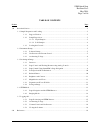
Sem quick start revision no.2 may 2014 page 2 of 16 table of contents section page 1.0 procedural section ............................................................................................................................................ 3 1.1 sample preparation and loading ...................
Page 7
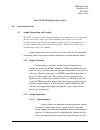
Sem quick start revision no.2 may 2014 page 3 of 16 zeiss vp-fe-sem quick start guide 1.0 procedural section 1.1 sample preparation and loading note: the cleanliness of the vacuum chamber is very important. It is encouraged that the investigator wear gloves while handling objects that will enter the...
Page 8
Sem quick start revision no.2 may 2014 page 4 of 16 are all fairly simple. Perhaps the most convenient instrument used to deposit spots on the support is the mechanical micropipette. The exact volume of the spot will naturally vary depending on the situation. It is advisable to test different sample...
Page 9
Sem quick start revision no.2 may 2014 page 5 of 16 1.2 instrument startup this section chronologically outlines the steps required to properly start up the instrument safely. 1.2.1 software startup the microscope is controlled entirely by the smartsem software suite developed by zeiss. The software...
Page 10
Sem quick start revision no.2 may 2014 page 6 of 16 the pressure of the sample chamber can be viewed under the vacuum tab on the right sidebar. Pressure units can be cycled through to the user’s preference by left-clicking the vacuum pressure status boxes. The microscope requires a vacuum below 0.00...
Page 11
Sem quick start revision no.2 may 2014 page 7 of 16 values. Compare these images with one another and make a mental note of their differences. 1.3 producing an image note: this section assumes that the operator is working in sem mode only and that no stem stage or detector will be used. The operator...
Page 12
Sem quick start revision no.2 may 2014 page 8 of 16 detector to be discussed is the stem detector, which is composed of both dark-field and bright-field components. The stem detector collects electrons that travel through the sample while mounted on stem- compatible carousel and support film. Since ...
Page 13
Sem quick start revision no.2 may 2014 page 9 of 16 operator is comfortable with. Be sure to select the tv detector channel while making changes to the working distance. The larger control stick on the right is responsible for moving the stage through three degrees of freedom. As depicted on the con...
Page 14
Sem quick star revision no.2 may 2014 page 10 of 16 1.3.4 scan speed and noise reduction the data coming from the currently selected detector channel on the instrument can be sampled and processed differently depending on the task at hand. The scan speed changes how fast the instrument samples data ...
Page 15
Sem quick start revision no.2 may 2014 page 11 of 16 keyboard . Once enabled, a small box will appear somewhere in the viewing window. Any changes made to the scene will only take place within the perimeter of this box. The location and size of the reduced raster window can be changed by clicking an...
Page 16
Sem quick start revision no.2 may 2014 page 12 of 16 1.3.8 stigmation and focus wobble. Producing well-focused images, especially at higher magnifications, requires the cross-section of the electron beam to be circular. The process of circularizing the electron beam is called stigmation and it is ca...
Page 17
Sem quick start revision no.2 may 2014 page 13 of 16 low and the noise reduction method should be set to line average. To freeze a frame, open the scanning tab on the sem control side-panel and select freeze on = end frame from the first dropdown menu in the noise reduction box. A red dot indicator ...
Page 18
Sem quick start revision no.2 may 2014 page 14 of 16 switching to the tv detector channel (section 1.3.1), open the stage points list tool and double click the stage point labelled $stem_safe_zone . The stage will move to an area in the chamber that is safe for detector arm insertion. Once the stage...
Page 19
Sem quick start revision no.2 may 2014 page 15 of 16 1.5 logging off finishing a session with the microscope is roughly the reverse of the startup process. The following sections will briefly outline the steps to safely log off the instrument. 1.5.1 stage positioning and stem detector the first step...
Page 20
Sem quick start revision no.2 may 2014 page 16 of 16 1.5.3 vacuum control and gas pressure note: if the instrument will be used again after a short period of time, or if the operator does not wish to vent the vacuum chamber, this step can be skipped. Venting the vacuum chamber can be accomplished by...
Page 22
Please make all necessary changes on the below label, detach or copy and return to the address in the upper left hand corner. If you do not wish to receive these reports check here ; detach, or copy this cover, and return to the address in the upper left hand corner. Office of research and developme...

